
To view a PDF version of this information, click here.
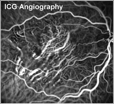
Indocyanine green angiography is a special test typically available only at major academic eye hospitals. It can provide helpful information about the deeper circulation of the eye in certain disease states. We are proud to offer this technology to our patients with particularly challenging diagnostic findings. This test is very similar in principle to fluorescein angiography, but instead of sodium fluorescein, a different dye called indocyanine green (ICG) is used. ICG dye produces a fluorescent signal at a different wavelength than fluorescein dye and also binds more strongly to proteins in the blood. ICG angiography has the advantage that it can provide information about deeper blood flow in vessels underneath the retina, called the choroid, and can be helpful in evaluating small "blisters" of fluid which can occur beneath the retina. Also, certain abnormalities like blood or pigment can block fluorescein dye, and in such cases, ICG may be able to locate sources of abnormal circulation and leakage because the signal penetrates deeper.
ICG angiography can take 10 to 20 minutes to perform and involves a small needle-stick. After dilation, your blood pressure will be measured. You will then be seated comfortably, and the doctor will inject a small amount of ICG dye into a vein on your arm or hand. At the same time, you will be instructed to place your chin in a chin-rest on the machine. You will then be instructed to look at a target while our skilled photographer takes digital photographs using the necessary filters. Your doctor will review the results with you in the examination room.
WARNING: You should notify our physicians and staff if you have a known allergy to penicillin, sulfa drugs, shellfish, or iodine before having this test.

