To view a PDF version of this information, click here.
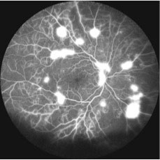
While a photograph of the retina provides a snapshot at one instant in time, an angiogram is a series of pictures that reveals the pattern of blood flow in the retina over a small period of time. Angiograms can be helpful for many conditions. For example, with an angiogram, your doctor will be better able to determine if the blood flow in the back of the eye is adequate, if there is inflammation within the blood vessels of the retina, if there is leakage of fluid and if so where that leakage comes from. These are only a few of the ways in which this important test can be helpful.
Wide field angiography allows up to a 200° view of the retina compared to 30-40° from traditional fundus photography. Furthermore it allows for better visualization of peripheral retinal diseases including neovascularization, nonperfusion, and previous laser treatment. This information can then be used to optimally treat the patient.
Sodium fluorescein is a dye used in fluorescein angiography (FA). The fluorescein dye is injected into the bloodstream (usually through a vein on the arm or hand). The dye will circulate in the blood vessels of the retina, and will give off a fluorescent color when light passed through special filters is focused on the retina. Dynamic pictures of the blood flow inside the eye are then taken.
FA can take 5 to 10 minutes to perform and involves a small needle-stick. After dilation, you will be seated comfortably, and the doctor will inject a small amount of fluorescein dye into a vein on your arm or hand. At the same time, you will be instructed to place your chin in a chin-rest on the machine. You will then be instructed to look at a target while our skilled photographer takes digital photographs using the necessary filters. Your doctor will review the results with you in the examination room.


