
To view a PDF version of this information, click here.
Vitrectomy surgery (or pars plana vitrectomy) is a common surgical procedure that is used for a wide number of conditions of the retina. These include: diabetic retinopathy, retinal detachment, macular pucker, macular hole and vitreous hemorrhage to name a few. The complexity of the procedure varies a great deal based on the underlying condition. For repair of retinal detachments, vitrectomy surgery may be performed alone or in combination with scleral buckle surgery.
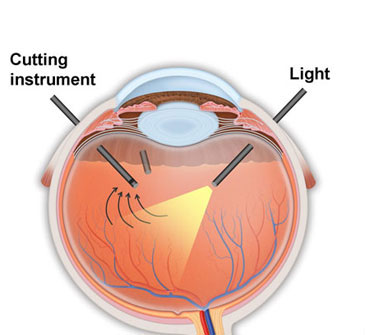
The surgery is performed by making three small incisions (about the size of a needle) through the white part of the eye known as the sclera. Through these incisions, fluid is introduced into the central portion of the eye while the vitreous jelly of the eye is removed by a rapid cutting instrument. The surgery is performed through an operating microscope. For more extensive work, other instruments such as microscissors or microforceps may also be introduced through these incisions to perform microsurgery near the retinal surface. Laser may also be performed through these incisions.
The latest major advance in vitrectomy surgery is the development of sutureless instruments and techniques. In traditional vitrectomy surgery, the wounds, although small, must still be closed with tiny absorbable sutures. With the latest instrumentation, the incisions are so small that they seal on their own and sutures are rarely, if ever, required. Although both techniques work well in appropriate cases, the newer sutureless approaches offer the added benefits of greater comfort following surgery and more rapid improvement in vision.
Before your procedure, your eye undergoing surgery will be dilated in the preoperative staging area by the nursing staff. There, you will meet with your surgeon as well as the anesthesiologist and operating room nursing staff. From there, you will be escorted to the operating room. Once in the operating room, you will have an IV started, EKG leads placed, and a blood pressure cuff applied to monitor you during the procedure. You will then be deeply sedated while your doctor administers your local anesthesia around your eye. You will gradually become more aware of your surroundings as your surgery is started, but you will remain comfortably and lightly sedated throughout the procedure. Many patients sleep throughout the entire process.
Once the retinal problem is repaired, it is occasionally necessary to replace the fluid of the eye with a gas bubble or oil bubble to help the retina heal in its proper location. This might require that you position your head in a specific manner after the procedure (sometimes even face down) for a determined period of time-sometimes even up to 1 to 2 weeks after surgery-particularly if your are having surgery for a macular hole. If a gas bubble is used, the duration of head positioning will be determined by your doctor during and after the surgery and discussed with you.
After your retinal problem is repaired, the incisions in the sclera and the surface layers of the eye may be closed with absorbable sutures that are thinner than a hair. Sutures may not be required at all if a sutureless approach is used. Antibiotics and steroids are injected over the surface of the eye to decrease inflammation and protect against infection after surgery. A patch is placed and then you are brought back to the recovery area where you will be observed for 1-2 hours prior to being released home. If sutures are used, they will initially feel a little scratchy and over 1 to 2 weeks, they will become softer and dissolve away.
Vision recovery after vitrectomy surgery depends largely on the underlying reason for the surgery. In some cases, surgery is performed in an attempt to prevent further vision loss, and in other cases, it is performed to improve the vision. Before recommending any surgical procedure, your doctor will discuss with you in detail the reason the surgery may be of benefit, the chances of vision loss, vision preservation, and vision gain with or without surgery, the risks of surgery, and the alternatives (if any) to surgery. If you are considering surgery, please be sure you have all your questions answered by our doctors and staff. If you leave and other questions arise, please feel free to contact us anytime.

