To view a PDF version of this information, click here.

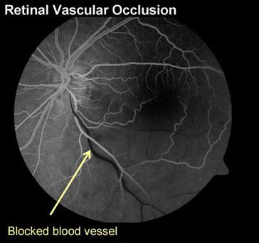
A simpler way to think of it is to imagine the blood vessels of the retina as plumbing. A blocked artery decreases the blood coming in, and a result, the area of retina downstream of that artery becomes pale, sick and damaged. A blocked vein slows the exit of blood and the system backs up, causing blood and fluid to spill over into nearby areas of retina. Blockages in either arteries or veins impair the circulation of the retina and can lead to painless vision loss. Just as with plumbing, blockages in larger blood vessels lead to more potential damage and vision loss. Because all or part of the retina may be affected, sometimes the vision loss may be only partial (that is, a part of the field of vision is lost) or total.
Blocked blood vessels, particularly veins, can lead to many of the same types of damage that are found in diabetic eye disease. The 3 most common reasons people with retinal vein occlusions lose vision are listed below:
Damaged blood vessels in and near the macula (the central part of the retina) can leak fluid and blood. This leakage of fluid can cause swelling and thickening of the macula, a condition known as macular edema. If we think of the eye as a camera and the macula is the film in the camera, then fluid leaking in the macula is like taking a picture with wet, warped film. If leakage persists for a long time, yellowish protein and fat deposits left behind by the fluid can build up inside the retina.
The treatment: The two most common treatments for macular edema are focal laser and medical injections. Laser is used to target the damaged, leaking blood vessels and stop the leakage. Injections of molecular medications into the eye are also used to stop fluid leakage from damaged blood vessels. Sometimes these two types of treatment are used together, and sometimes repeat treatment sessions are needed.
When blood vessel blockage and damage is more severe, the retina tries to compensate by growing new blood vessels. This is the retina's natural response to poor blood flow. Unfortunately, rather than helping the situation, these new blood vessels make things worse because they are abnormal and fragile and they rupture and bleed very easily.
If they continue to grow, abnormal blood vessels in the eye can bleed into the vitreous cavity causing severe floaters and severe vision loss.
The treatment: The best time to treat abnormal blood vessel growth is before bleeding occurs. Panretinal laser is applied to the peripheral retina in the area of the blockage, where the blood flow is the poorest. This slows the growth of new abnormal blood vessels and usually causes existing abnormal blood vessels to shrink and wither away. Multiple sessions of laser may be required.
If there is severe bleeding inside the eye, then laser treatment cannot be performed because the blood blocks the doctor's view into the eye, making it unsafe to use laser. If this happens, waiting and keeping the head elevated at all times often helps the loose blood in the eye settle down enough to enable the doctor to apply laser on a future visit. If this blood does not settle down or clear, then vitrectomy surgery may be needed to clean the blood out and restore vision.
Sometimes, the blood vessel damage is so severe that there is not enough blood flow to nourish the macula, the central part of the retina. The macula gradually becomes starved for nutrients and oxygen and is not able to function well, resulting in vision loss. Unfortunately, the blood vessels of the macula are too tiny to be repaired and if this problem occurs it cannot be reversed. But even if the blood flow is not adequate, treating the other problems above can still maximize what vision there is in the eye.
Retina vascular occlusions can be associated with other conditions of the eye (glaucoma) or the body (high blood pressure, high cholesterol, clotting disorders, inflammatory blood vessel diseases, etc.). If you are diagnosed with a retinal vascular occlusion, it is important, in addition to seeing your Los Angeles ophthalmologist, to follow up with your primary doctor. Sometimes, vascular occlusions of the retina may be an early warning sign of an even more serious risk, such as a stroke. As part of your healthcare team, your retina doctor will discuss these risks with you and will coordinate your care, and any necessary blood testing, with your primary doctor.

